Chick Embryo Microangiography
Chick Embryo Microangiography
MRI of Chick Embryos
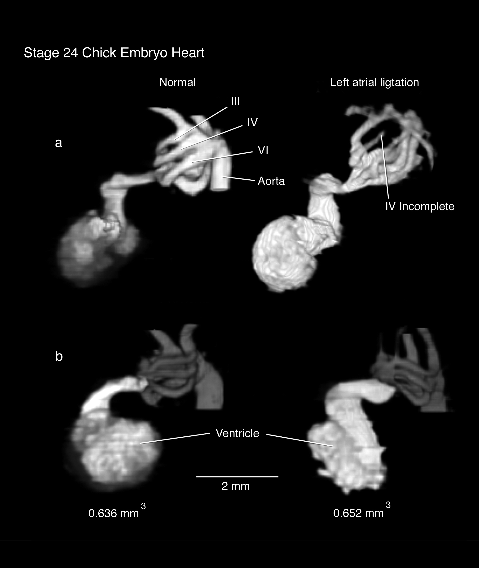
MRI of Stage 24 Embryo Ventricle and Aortic Arches
MRI of Stage 24 Embryo Heart Outflow Tracts,
Normal and After Atrial Ligation
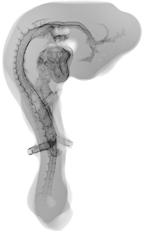
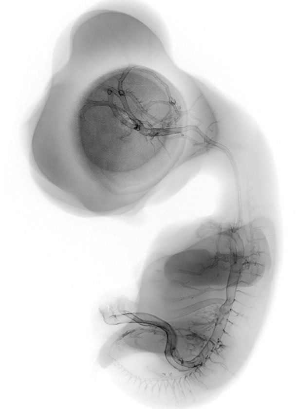
MR microscopy of chick embryo vasculature was performed by adapting imaging methods developed at the Center for In-vivo Microscopy at Duke University* and perfusion protocols developed for mouse microangiography (x-rays) by Eric L. Effmann**.
Fertilized Arbor Acre eggs were incubated at 37.5 ºC and 55% humidity with twice daily rotation until the time of vascular perfusion. Windows in the shell were created directly over the embryos for perfusion. Perfusion was performed through a four-channel Gilson peristaltic pump and finely drawn glass micropipette (25-75 micrometer tip diameter) at flow rates of approximately 2-3 microliters per minute. A small tear was made into a vitelline or allantoic artery and vein (depending on embryonic age). The vein was cannulated with the micropipette and perfused with 37ºC fixative (2% glutaraldehyde and 1%formaldehyde in 300 mOsm phosphate buffer supplemented with 0.015% Brilliant Blue dye to visually track perfusion progress). After the fixative had replaced the embryonic blood, the artery was perfused with a warmed phosphate buffered solution, followed by perfusion with an MRI contrast agent (0.003-mol/L gadolinium chloride in a 10% agarose solution). The cannulated vessels were ligated and the embryos immersion fixed in cold perfusion fixative (minus the blue dye) immediately following the gadolinium perfusion for 1 week. Fixed embryos were placed into a plastic vial and surrounded by a warmed solution of 10% agarose and allowed to cool and solidify prior MR imaging.
Magnetic resonance imaging was performed at 9.4 Tesla with a 1 cm solenoid imaging coil using a three-dimensional spin-echo pulse sequence with the following approximate parameters (depending on embryo age/size): repetition time (TR) = 198 ms, echo time (TE) = 7 ms, number of excitations per view (nex) = 2, flip angle = 90, with fields of view and slice numbers resulting in isotropic voxels, and total scan times on the order of 3 hrs.
* Smith BR, Effmann EL, Johnson GA. MR microscopy of chick embryo vasculature. J Magn Reson Imaging. 1992 Mar-Apr;2(2):237-40.
** Effmann EL, Whitman S, Pexieder T. Stereo microangiography in embryologic and teratologic investigation. Teratology. 1986 Aug;34(1):103-12.
© 2026.
Chick Embryo Microangiography by Brad Smith is licensed under CC BY-NC-SA 4.0![]()
![]()
![]()
![]()